
SkinRehab™ · Clinical Skin Checks & Mapping
Early detection is everything.
Skin cancer screening combines clinical observation, dermatoscopy, palpation and a detailed medical history to form the foundation of your assessment. Every skin check is performed with a dermatoscope, at every appointment, so nothing on your skin goes unexamined.
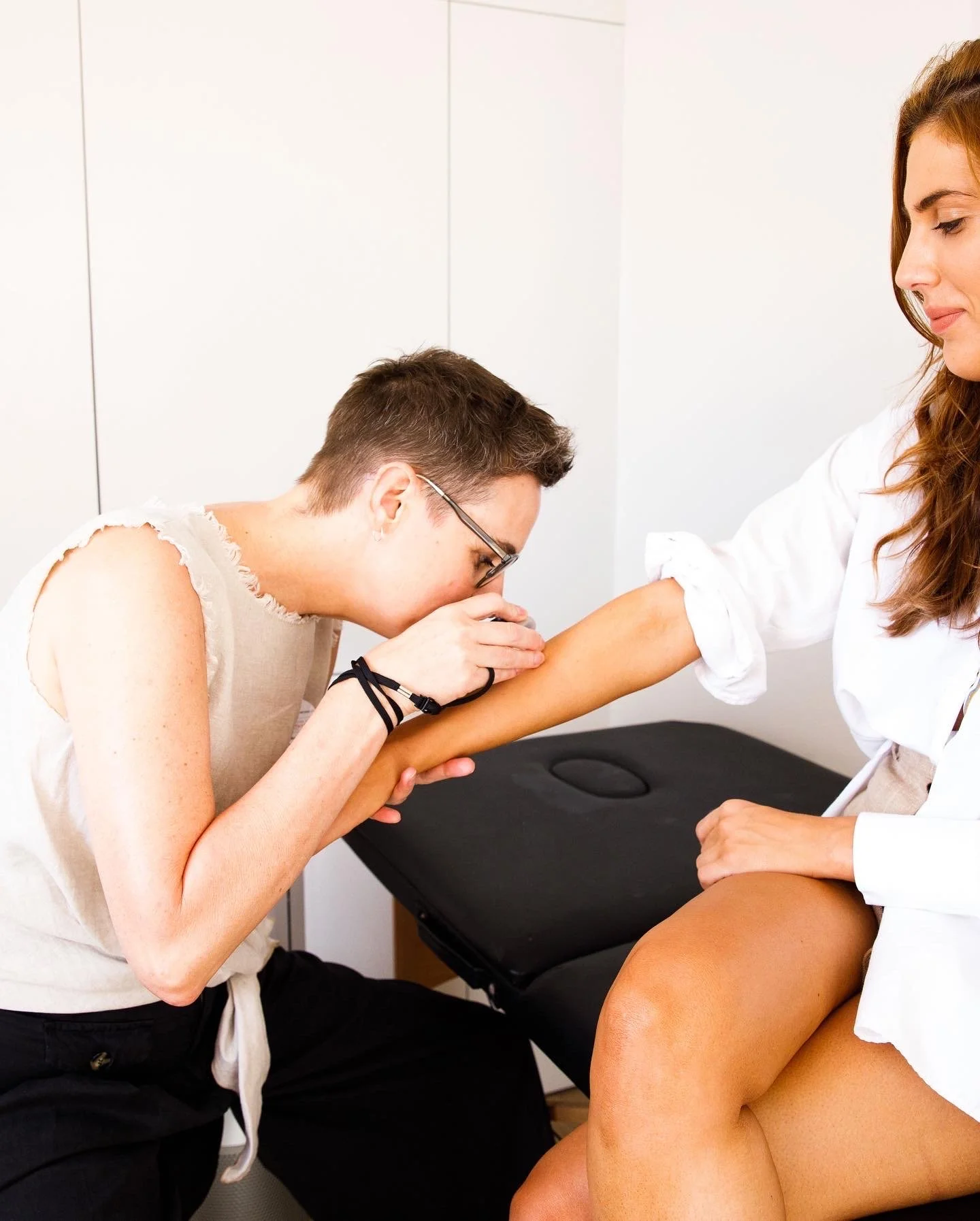
Clinical screeningA partnership in surveillance.
Between appointments
Your assessment is strengthened by your participation. Regularly checking your own skin between annual visits sharpens early detection, because you are the first person positioned to notice a new or changing lesion. Anything you observe becomes part of the clinical picture at your next appointment.
Photographic documentation
Digital images support the assessment of benign and malignant conditions, but they are never a substitute for clinical confirmation. Where photographic documentation is needed, consent and release are addressed with you during your appointment.
The instrumentWhat is dermatoscopy?
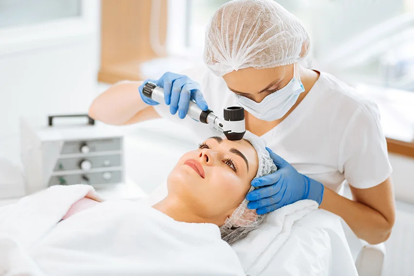
Dermatoscopy, also called dermoscopy, is the analysis of the skin, and in particular its lesions, with a dermatoscope: a hand-held instrument that combines magnification and illumination to reveal the intricate patterns of benign and malignant lesions, and the vasculature of inflammatory skin conditions.
All skin checks and Skin Health Assessments are performed with dermatoscopy. This gives the most complete analysis of your overall skin health, your sun exposure status, and the primary presentation of any skin conditions. It is particularly valuable when determining treatment strategies for restorative therapies, and when recommending corrective skincare and sun protection moving forward.
Total body photographyWhat is mole mapping?
01
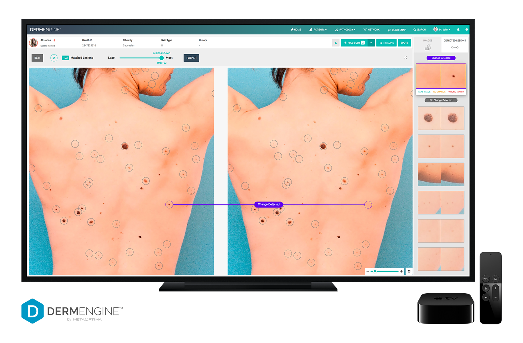
Total body photography, widely known as mole mapping, is the process of digitally imaging lesions and skin conditions with specialised medical technology, then plotting them across the total surface area of the skin. Mapping serves two purposes: detecting slow change or progression within individual lesions, and establishing the baseline morphology of every lesion across your body.
02
The mapping software combines dermatoscopy with artificial intelligence to support early detection and ongoing monitoring of the skin. Early detection of change is the single most important factor in the prognosis and survival of melanoma and skin cancer.
03
Monitoring with innovative medical technology delivers something no self-examination can: the assurance of knowing your skin is under structured, documented surveillance. It is this assurance that underpins The Dermal Health Alliance's mantra of early detection and prevention.
Who benefits most
Complex cases requiring structured monitoring and specialist referral.
Individuals at high risk of skin cancer or melanoma.
Anyone with multiple moles, where baseline mapping makes change visible.
Anyone seeking documented, longitudinal surveillance of their skin.
Total body photographyEvery lesion is plotted and matched across visits. The software compares imaging over time and flags any lesion where change is detected.
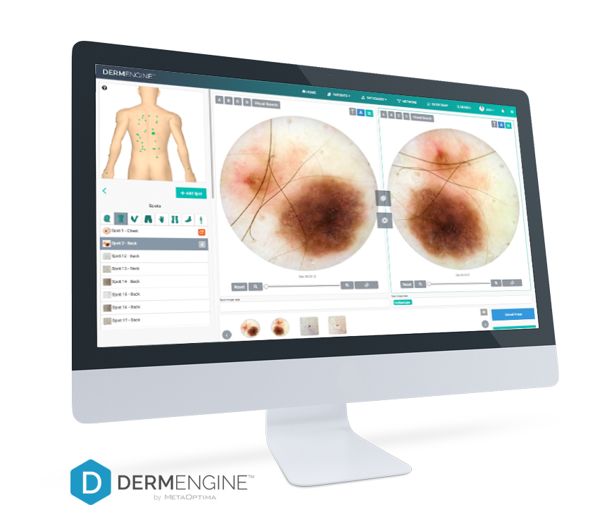
Individual lesion monitoringLesions of interest are imaged dermoscopically, mapped to their body site and tracked against dated baselines.
-
The DermEngine Team. (2019, September 23). The future of dermatology: AI implementation in real-life scenarios [Web log post]. Retrieved from http://www-DermEngine-com.cdn.ampproject.org
Kittler, H. (2008). Early recognition at last. Archives of Dermatology, 144(4). doi:10.1001/archderm.144.4.533
Kittler, H. (2012). The order of things and the classification of melanoma. Dermatology Practical & Conceptual, 2(4). doi:10.5826/dpc.0204a01
Madigan, L. M., Treyger, G., & Kohen, L. L. (2016). Compliance with serial dermoscopic monitoring: An academic perspective. Journal of the American Academy of Dermatology, 75(6), 1171-1175. doi:10.1016/j.jaad.2016.07.012
Madnani, N. (2012). Dermoscopy in Clinical Practice: Using Your iPhone for Dermoscopy. Dermoscopy and Trichoscopy in Diseases of the Brown Skin: Atlas and Short Text, 232-232. doi:10.5005/jp/books/11693_20
Rosendahl, C., Cameron, A., Tschandl, P., Bulinska, A., Zalaudek, I., & Kittler, H. (2014). Prediction without Pigment: a decision algorithm for non-pigmented skin malignancy. Dermatology Practical & Conceptual. doi:10.5826/dpc.0401a09
Sharma, P., Vleugels, R. A., & Nambudiri, V. E. (2019). Augmented Reality in Dermatology: AR we ready for it? Journal of the American Academy of Dermatology. doi:10.1016/j.jaad.2019.07.008
Singh, N., & Gupta, S. K. (2018). Recent advancement in the early detection of melanoma using computerized tools: An image analysis perspective. Skin Research and Technology, 25(2), 129-141. doi:10.1111/srt.12622
Tschandl, P., Argenziano, G., Razmara, M., & Yap, J. (2018). Diagnostic accuracy of content‐based dermatoscopic image retrieval with deep classification features. British Journal of Dermatology, 181(1), 155-165. doi:10.1111/bjd.17189
Zalaudek, I., Argenziano, G., & Giacomel, J. (2016). Dermatoscopy of Non-Pigmented Skin Tumors: Pink - Think - Blink. Boca Raton, FL: CRC Press.
Early detection & prevention.
Every skin check is performed with dermatoscopy by a credentialed Dermatoscopist and Melanographer. Book your clinical skin check and put your skin under expert surveillance.